
Figure 2. Weight gain is positively correlated with perilymphatic inflammation .
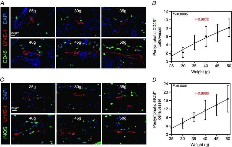
A, representative photomicrographs of hindlimb skin immunofluorescence staining localizing leukocytes (CD45+; green) with lymphatic vessels (LYVE‐1+; red) and nuclei (DAPI; blue). Note the increase in perilymphatic leukocytes as weight increases. B, quantification of perilymphatic CD45+ cells (cells located within 50 μm of lymphatic vessels) for each weight point (n = 5 animals per group with four HPFs per animal; Pearson's correlation: r = 0.9872, P = 0.0002). C, representative photomicrographs of hindlimb skin immunofluorescence staining localizing iNOS (iNOS+; green) with lymphatic vessels (LYVE‐1+; red) and nuclei (DAPI; blue). Note the increase in perilymphatic iNOS+ cells as weight increases. D, quantification of perilymphatic iNOS+ cells (cells located within 50 μm of lymphatic vessels) for each weight point (n = 5 animals per group with four HPFs per animal; Pearson's correlation: r = 0.9986, P < 0.0001).