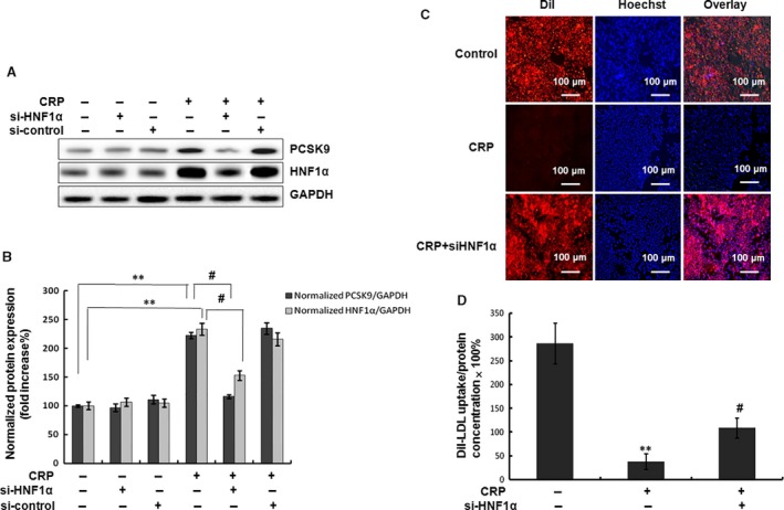
Figure 6.

Inhibition of HNF1α attenuated the PCSK9 expression and LDL uptake by CRP in HepG2 cells. (A) (B) Western blot analyses of HNF1α and PCSK9 in HepG2 cells transfected with siRNA‐HNF1α (80 nM) and then treated with CRP (10 μg/ml) for 24 hrs. (C) Representative fluorescence microscopy images of cell‐associated Dil‐LDL (red), Hoechst‐stained nuclei (blue) and the overlay. (D) Fluorescence of isopropanol‐extracted Dil (520–570 nm, normalized to the cell protein). Data were presented as mean ± SEM (n = 3). Significance: *P < 0.05, **P < 0.01 versus control; # P < 0.05, ## P < 0.01 versus the CRP‐treated group. CRP, C‐reactive protein; PCSK9, pro‐protein convertase subtilisin/kexin type 9; HNF, hepatocyte nuclear factor.
