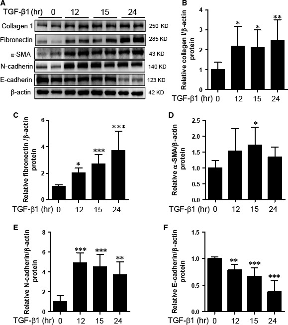
Figure 1.

Extracellular matrix (ECM) proteins and epithelial‐mesenchymal transition (EMT) markers are induced by TGF‐β1. HK‐2 cells were incubated with TGF‐β1 (5 ng/ml) for the indicated time points. (A) Representative immunoblots. (B–F) Fibronectin, collagen type I, α‐SMA, N‐cadherin and E‐cadherin protein expression was quantified by densitometry. Data are expressed as mean ± SD of six independent experiments. *P < 0.05, **P < 0.01, and ***P < 0.001 compared with untreated cells.
