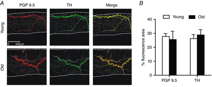
Figure 6. Sympathetic innervation of GM feed arteries for young and old mice .

A, representative immunofluorescent staining (maximum Z‐stack projection of confocal image slices) of all perivascular nerves (protein gene product 9.5, PGP9.5) and of sympathetic nerves (tyrosine hydroxylase, TH) in GM feed arteries of young and old mice. Vessel edges indicated by dotted lines. B, innervation per vessel surface area (% fluorescence area; see Methods) for PGP9.5 and TH was not different between FAs of young and old mice. Summary data are means ± SEM, n = 4 per age group.
