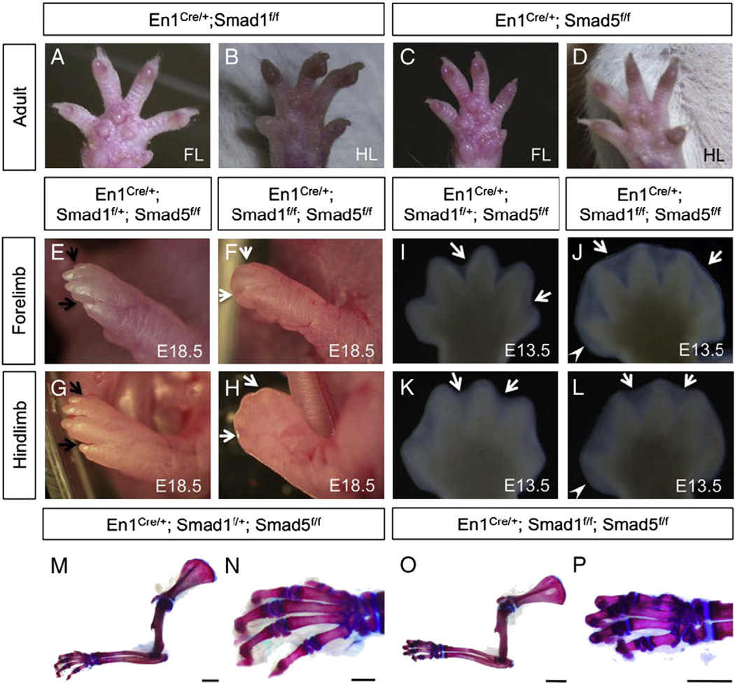
Fig. 2.
Inactivation of Smad1 and Smad5 in the AER and ventral ectoderm by En1Cre/+ knock-in allele resulted in syndactyly and retention of interdigital tissue. Inactivation of either Smad1 (A, B) or Smad5 (C, D) in the AER and ventral ectoderm did not result in visible phenotypes in both forelimbs (FL) and hindlimbs (HL) of the individuals at adult stage. (E–H) Fore- and hindlimb from the control (E, G) and Smad1/5 mutant fetuses (F, H) at E18.5. Black arrows indicate separated digits in the control, while white arrows indicate the retained interdigital tissue in the mutants. (I–L) The defect in interdigital tissue regression was observed in both fore- and hindlimbs of the Smad1/5 mutants starting from E13.5. Syndactyly (webbing) is observed in the double mutants (J, L). White arrows indicate interdigital tissue. White arrowheads indicate that the posterior end of the autopod of Smad1/5 mutant is broadened at E13.5. (M–P) Skeletal preparations of the forelimbs from the control (M) and Smad1/5 mutants (O) at postnatal day 10. Higher magnification view of the autopod of the controls (N) and the mutants (P). Inactivation of Smad1 and Smad5 in the AER and ventral ectoderm did not result in skeletal defects in the autopod and along proximal–distal axis. A–D are the ventral views of the limb with anterior to the right. E–L are dorsal views of the limb plates with anterior to the right. Scale bars: M, O=5 mm; N, P=500 µm.

