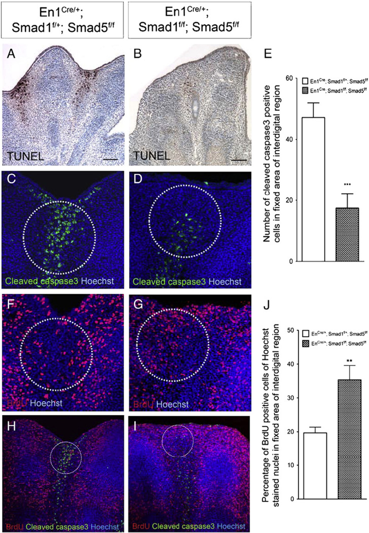
Fig. 3.
Smad1/Smad5 signaling in the AER and ventral ectoderm is required for regulating cell death and cell proliferation in interdigital regions. (A, B) TUNEL assay and (C, D) immunofluorescence detecting cleaved caspase3 to measure cell death in E13.5 forelimbs. Inactivation of Smad1 and Smad5 resulted in a decrease in cell death. (E) Quantification of the number of anti-cleaved caspase3 positive cells demonstrated that there was a significantly decrease in the number of cells undergoing cell death in the Smad1/5 mutant limbs (***p<0.005). (F, G) Smad1/5 signaling in the AER and ventral ectoderm is required for regulating cell proliferation in the underlying mesenchyme. BrdU labeling assay showed that inactivation of Smad1 and Smad5 resulted in an increase in cell proliferation in distal interdigital mesenchymal regions. (H, I) Cleaved caspase3 and BrdU co-immunostaining indicated that distal interdigital areas (white circle) of Smad1/5 mutant are abundant in BrdU positive cells. (J) Quantification of the percentage of BrdU positive cells with Hoechst stained nuclei demonstrated that there was a significant increase in the percentage of proliferating cells in the distal interdigital areas of the Smad1/5 mutant limbs (**p<0.005). Scale bars: A–B=100 µm.

