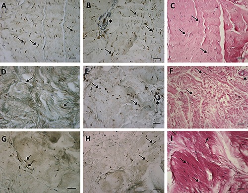
Figure 1.

ERα (A,D,G) expression, RXFP1 (B,E,H) expression and H&E staining (C,F,I) of paraffin sections from samples collected from three different districts of human fascia of three women patients: the rectus sheath of abdomen (A-C), the crural fascia of the leg (D-F), the fascia lata of the thigh (G-I). Arrows indicate some fibroblasts (in A,D,G positive for ERα and in B,E,H positive for RXFP1); asterisks show blood vessels, positive for RXFP1 in B,E. Scale bars: 50 μm.
