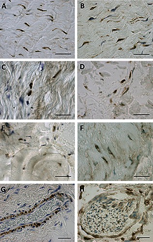
Figure 2.

ERα (A,C,E) and RXFP1 (B,D,F) expression of paraffin sections of the rectus sheath of the abdomen (A,B), the crural fascia of the leg (C,D), and the fascia lata of the thigh (E,F). G and H show a RXFP1 positive blood vessel and nerve, respectively. Scale bars: 50 μm.
