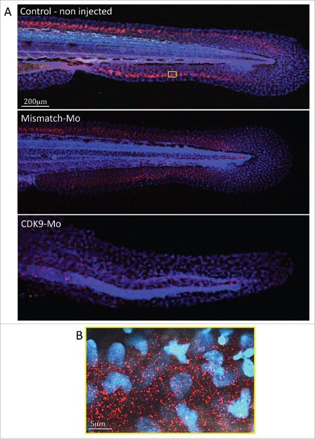
Figure 6.

Immunostaining for CDK9 in whole larvae. (A) Confocal images of zebrafish embryo (Wik, wild type) 72 hpf control non-injected (above), injected with mismatch morpholino (middle) or CDK9-targeted morpholino (low) and immunostained with anti-CDK9 antibody, in red, and counterstained with DAPI. The staining shows diffuse presence of CDK9, in both nucleus and cytoplasm. The small yellow boxed area in a control non injected larva is shown at higher magnification in (B).
