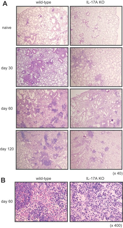
Figure 2.

Reduction of the lesion size in the lungs of IL‐17A KO mice after M. tuberculosis infection. Wild‐type C57BL/6 or IL‐17A KO mice were inoculated i.t. with M. tuberculosis H37Rv. The mice were sacrificed 30, 60, and 120 days after infection, and formalin‐fixed sections were stained with hematoxylin and eosin. Representative lung tissue specimens from the wild‐type C57BL/6 mice (left panel) and the IL‐17A KO mice (right panel) are shown. Magnification, ×40 (A), ×400 (B).
