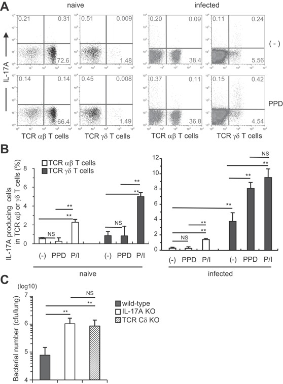
Figure 4.

TCR γδ+ T cells were the major IL‐17A‐producing cells in the lungs of M. tuberculosis‐infected mice. Wild‐type C57BL/6 mice were inoculated i.t. with M. tuberculosis H37Rv or left untreated (A and B). The PIF cells (5 × 10⁵ cells) were prepared on day 60, and were cultured with PPD (5 μg/ml) in the presence of naive spleen antigen‐presenting cells (1 × 10⁵ cells) for 18 h at 37°C, and with GolgiPlug for the last 6 h. The cells were also stimulated with PMA and ionomycin. After the culture, the cells were surface stained with FITC‐CD3e, PerCP‐Cy5.5, or APC‐conjugated anti‐TCR Cβ and APC‐conjugated TCR Cδ mAbs. Surface‐stained cells were subjected to intercellular cytokine staining with a PE‐conjugated anti‐IL‐17A mAb. The samples were analyzed by FCM (A, naïve; B, infected). Wild‐type C57BL/6, IL‐17A KO, and TCR Cδ KO mice were inoculated i.t. with 1 × 10³ CFU of M. tuberculosis H37Rv (C), and the CFU in the lungs was determined on day 60 after the infection. The statistical analysis was performed with ANOVA. Asterisks (*) indicate significant difference between two groups.
