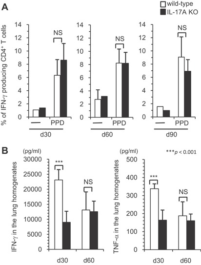
Figure 6.

The Th1 immune response in the early phase against M. tuberculosis infection. Wild‐type C57BL/6 or IL‐17A KO mice were inoculated i.t. with M. tuberculosis H37Rv. Mice were sacrificed 30, 60, and 90 days after infection, and the lung lymphocytes were stimulated with PPD, and the percentages of IFN‐γ producing CD4+ cells in the CD3+ T cell population were determined using FCM (A). In some experiments, the concentrations of IFN‐γ in the supernatants of the lung lymphocytes were determined by an ELISA using the DuoSet ELISA development kit, according to the manufacturer's protocol (B). The statistical analysis was performed with Student's t‐test. Asterisks (*) indicate significant difference between two groups.
