
Figure 1. Magnetic resonance imaging (MRI) protocol .
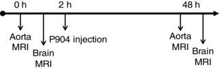
A pre‐contrast imaging (0 h) protocol was performed on the aorta, followed immediately by brain imaging with gadolinium injection. An ultrasmall iron oxide nanoparticles (USPIO) contrast agent, P904 (Guerbet, Aulnay‐sous‐Bois, France), was then injected. Forty‐eight hours later (48 h), an identical post‐USPIO aortic imaging protocol was performed for assessment of inflammation. This was followed immediately by brain inflammation imaging.