
Figure 5. Aorta magnetic resonance imaging of old and young untrained and old trained ApoE−/− mice .
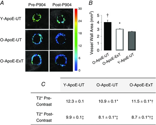
A, pre‐ and post‐USPIO T2* maps of the ascending aorta in a young untrained (Y‐ApoE‐UT), an old untrained (O‐ApoE‐UT) and an old trained (O‐ApoE‐Ext) ApoE−/− mouse. B, ascending aorta vessel wall area measurements. C, vessel wall pre‐ and post‐USPIO T2* measurements. In O‐ApoE‐UT mice, vessel wall area is significantly larger, and pre‐ and post‐USPIO T2* significantly lower, representative of advanced and complex atherosclerotic lesions with inflammatory activity. *Significantly different (P < 0.05) from old ApoE−/− mice (O‐ApoE‐UT). †Significantly different (P < 0.05) from young ApoE−/− mice (Y‐ApoE‐UT). ‡Significantly different (P < 0.05) from pre‐contrast.