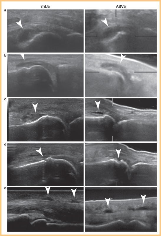
Fig. 2.

Representative corresponding images of synovitis, erosions and tenosynovitis (arrows) detected by mUS and ABVS. mUS and ABVS images were acquired with 18 MHz and 11 MHz probes, respectively. a MCP 3 joint with grade 2 synovitis. b MTP 2 joint with grade 2 synovitis. c MCP 3 joint with grade 1 synovitis. d MCP 3 joint with erosions. e Index finger tenosynovitis of the flexor tendon.
