
Figure 1.
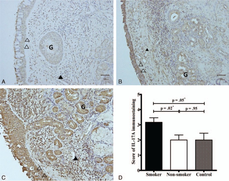
Immunohistochemistry findings of evaluation of interleukin (IL)-17Aprotein expression in nasal tissues. The intensity of immunostaining was scored as weak: <35% (A, scored 1); moderate, 35–70% (B, scored 3); or strong, >70% (C, scored 5) based on the proportion of positively stained cells. The IL-17A expression levels in nasal tissues of smokers were higher compared to those in the nasal tissues of nonsmokers and control subjects (D). ∗Significance was considered at P < 0.05, analyzed by the Mann–Whitney U test. Cells were observed at a magnification of 200×. Open head, epithelium; closed head, endothelium; arrowhead, inflammatory cells; G, mucus gland. IL = interleukin.