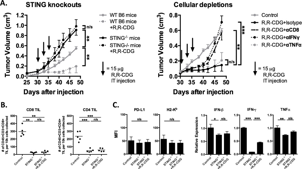
Figure 5. Control of MOC1 tumors following CDN treatment is STING and CD8+ T lymphocyte–dependent.
A, WT or Tmem173gt/J STING-deficient B6 mice were injected with 2 × 10⁶ MOC1 cells and established tumors were treated with intratumoral R,R-CDG (15 µg/injection q 3 days × 3, n = 5–7 mice/group, left panel). Black arrows indicate individual injections. Right panel, established MOC1 tumors were treated with intratumoral R,R-CDG (15 µg/injection q 3 days × 3) along with systemic administration of CD8+ T cell–, IFNγ-, or TNFα-depleting antibodies (200 µg/injection IP twice weekly). Depleting antibodies were started 48 hours before R,R-CDG treatment. Control and STING-deficient mouse tissues were harvested 48 hours after the last R,R-CDG treatment and analyzed with flow cytometry and RT-PCR. B, quantification of CD8+ and CD4+ TILs in STING-deficient mice (left panels). C, flow cytometric quantification of PD-L1 and H2-Kb on live CD45.2–CD31– tumor cells (left panels) and RT-PCR–based determination of IFNβ, IFNγ, and TNFα expression relative to control (right panels, n = 3–5 tumors/group). *, P < 0.05; **, P < 0.01; ***; P < 0.001; ANOVA. n/s, not significant.

