Figure 4.
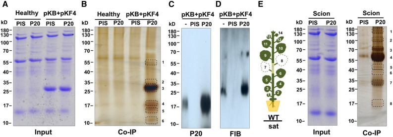
Identification of P20-Interacting Protein Complex from Grafting N. benthamiana Leaves with HV-Dependent or -Independent SatBaMV Infection by Co-IP.
(A) Coomassie blue staining of total protein extracted from leaves of healthy or BaMV (pKB) + satBaMV (pKF4) (Liou et al., 2013) coinfected N. benthamiana at 7 DPI.
(B) Co-IP protein complexes by preimmune IgG (PIS) or anti-P20 IgG (P20) were separated by SDS-PAGE. Protein bands were visualized by silver staining; frames indicate protein bands excised for LC-MS/MS protein identification. The gel is representative of three independent experiments.
(C) and (D) Detection of P20 and fibrillarin in co-IP complex from anti-P20 IgG or PIS antibody. Input (−) and complex were separated by SDS-PAGE followed by immunoblot analyses with anti-P20 (C) or anti-FIB IgG (D).
(E) HV-independent grafting experiments are illustrated in the left as in Figure 2B. Total protein was extracted from wild-type scion leaves after grafting at 15 DAG. Co-IP and protein analysis were performed as in (B).

