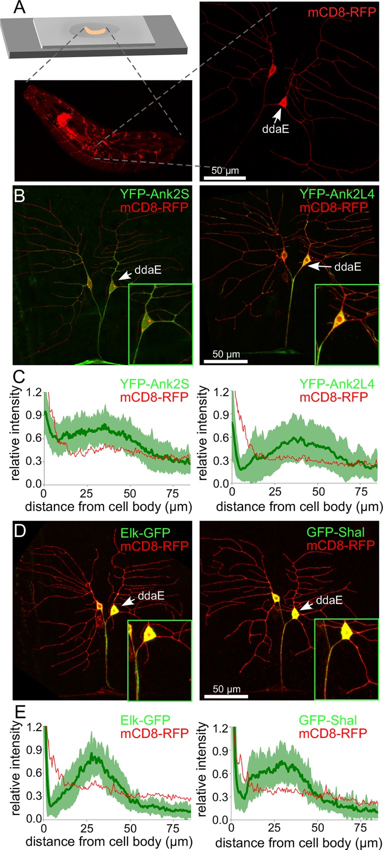
Fig 6. Enrichment of a Drosophila Ank2L polypeptide and two tagged ion channels in the proximal axon.
A. The strategy to image tagged proteins in whole Drosophila larvae is shown. Larvae are placed on a drop of agarose dried onto a slide and held in place by a coverslip taped onto the slide to prevent movement (top left). Neurons on the dorsal side of the animal can be visualized through the cuticle (lower left); the dendrites, cell body and proximal axon of the ddaE neuron are all easily visible (right). B and D. YFP-Ank2S, YFP-Ank2L4, Elk-GFP and GFP-Shal were expressed in class I neurons using 221-Gal4. mCD8-RFP was also expressed to outline the cells. The two dorsal class I neurons are shown, with ddaE at the right and ddaD on the left. Insets show the cell body, proximal axons and proximal dendrites of ddaE. C and E. To quantitatively assess marker distribution, the fluorescence intensities were measured from the cell body along the axon. Averages from 30 cells are shown for each marker. For the Ank2 constructs, and tagged Elk and Shal, the standard deviation is shown as light green around the dark green average line. For mCD8-RFP only the average is shown for comparison.

