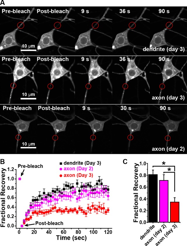
Fig 7. Diffusion in the plasma membrane is restricted at the base of the axon compared to the base of the dendrite.
A. Example images of dendrite and axon FRAP experiments at two different larval ages are shown. mCD8-RFP was expressed in class I dendritic arborization neurons. Bleaching was performed in the ddaE neuron either at the base of the comb-like dendrite or the base of the axon. The red circle indicates the bleach area. B. The average recovery of fluorescence into the bleach areas as shown in A is plotted on the graph; averages were calculated from 14 cells for the dendrite, 17 for axon 2 day and 13 for axon 3 day. Each cell was in a different animal. The error bars show the standard error of the mean. C. Bars show the recovery plateau (mean ± SEM) quantitated by averaging recovery values between 110 and 120 ms. The asterisk indicates significant difference (P < 0.01, t-test).

