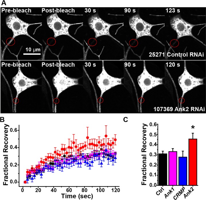
Fig 9. RNAi targeting Ank2L reduces the axonal diffusion barrier.
A. mCD8-GFP was expressed in class I neurons, and proximal axons of ddaE neurons were photobleached. Example images of a control neuron are shown in the top row, and Ank2 RNAi neurons below. B. Quantitation of FRAP experiments in different genetic backgrounds is compiled in the graph. The average recovery is shown, with standard error shown as error bars. Number of cells tested for each genotype were: control-24, Ank2-19, Ank-12, CRMP-9. C. The recovery plateau (mean ± SEM) was quantitated by averaging recovery values between 110 and 120 ms. The asterisk indicates significant difference (p < 0.02, t-test).

