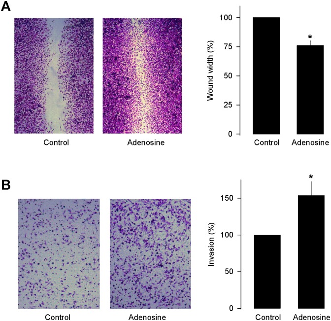
Fig 2. Effect of Ado on migration of MDA-MB-231 cells.
(A) Wound-healing migration assay for MDA-MB-231 cells in the absence and presence of 50 μM Ado (left panel). The percentage of migration in the transwell assay is shown in right panel. (B) The cell migration was assessed by the transwell migration assay in MDA-MB-231 cells incubated in the absence (control) or presence of 50 μM Ado. Shown are representative micrographs of the migrated cells stained with crystal violet (left panel) and the percentage of migrated cell (right panel). The results represent the means ± SE of three independent experiments performed in triplicate. Asterisks denote significant differences (p<0.05) compared to control.

