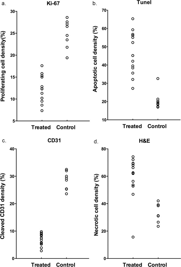
Fig 5. Histopathological analysis of tumors of control and treated group.
Histopathological analysis of (a) proliferating cell density (Ki67 staining), (b) apoptotic cell density (TUNEL staining), (c) microvessel density (CD31 staining) and (d) necrosis fraction (H&E staining) of the treated group and control group on day 7 after treatment.

