. 2014 Dec 31;7(4):1140. doi: 10.4022/jafib.1140

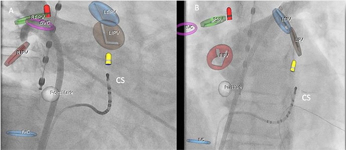
Figure 7. Snapshot of two simultaneously displayed MediGuideĀ® screens showing the left atrial mapping process during catheter ablation of atrial fibrillation. Sensors at the tip of various catheters are projected in real-time on pre-recorded loops (red tip for mapping catheter, yellow tip for coronary sinus catheter). Left (right anterior oblique view): Background movie showing semi-selective pulmonary vein angiography of the right upper pulmonary vein. Right (left anterior oblique view): Pre-recorded semi-selective left upper pulmonary vein angiography was chosen as the background for non-fluoroscopic sensor tracking. Various markers were set with the MediGuideĀ® system (RSPV, RIPV, LSPV, and LIPV for ostia of right/left superior/inferior pulmonary veins; SVC, IVC for superior/inferior caval vein; PointMark for oval fossa). At the time of recording the background loops, the transseptal sheath, coronary sinus catheter (CS), and esophageal probe were already in place.

Figure 7.