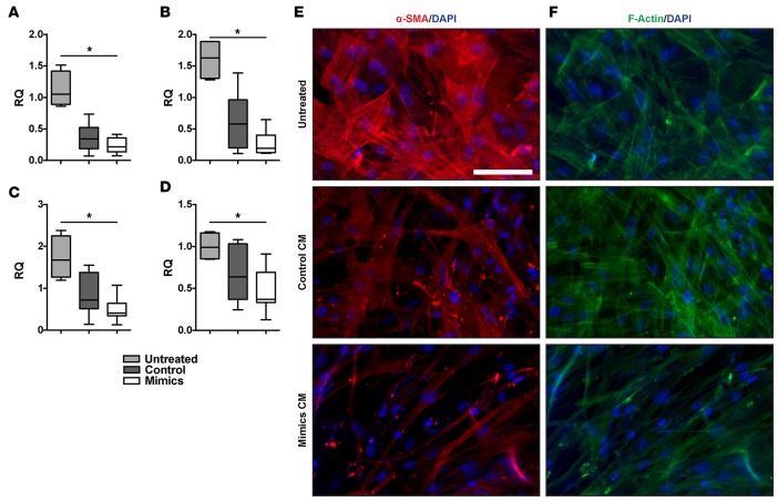
Figure 7. Epithelial suppression of fibroblasts is augmented by miR-323a-3p.
Conditioned medium from 16HBE14o- cells expressing either miR-323a-3p or control were incubated with primary human fibroblasts and compared with untreated fibroblasts. Expression of (A) α-SMA (ACTA2), (B) type I collagen (COL1A1), (C) type III collagen (COL3A1), and (D) fibronectin (FN1) was measured by PCR in fibroblasts. n = 4, *P < 0.005. (E) Immunofluorescence staining for α-SMA and (F) phalloidin in fibroblasts treated with epithelial cell–conditioned medium. Scale bar: 100 μm. Data are presented as whisker-box plots. The central horizontal bars indicate the medians, boxes indicate 25th to 75th percentiles, and whiskers indicate 1.5 times the interquartile range from the bottom and the top of the box. Statistical analysis in this figure was performed with a 1-way ANOVA. RQ, relative quantification.

