Fig. 3.
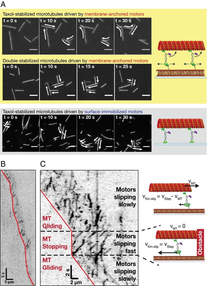
Membrane-anchored kinesin-1 motors slip in the lipid bilayer, while propelling a microtubule. (A) Time-lapse images for microtubules driven by membrane-anchored motors (Upper) and surface-immobilized motors (Lower) with schematic experimental setups on the Right. (Scale bar, 5 µm.) The arrows indicate the transport directions of the gliding microtubules. Microtubules propelled by membrane-anchored motors do not cross each other in contrast to microtubules driven by surface-immobilized motors. (B and C) Representative kymographs (inverted contrast) showing the movement of individual rKin430-SBP-GFP motors (dark signals) while propelling microtubules at low motor density (B) and high motor density (C). The red lines mark the trailing ends of the microtubules as guides to the eye. In C, a gliding microtubule collides with another passing microtubule and is temporarily stalled (also shown in the schematics on the Right) until the other microtubule glides away.

