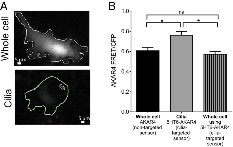
Fig. 1.
(A) Images of mouse embryonic fibroblasts expressing the PKA sensor AKAR in the whole cell or cilia (5HT6-AKAR4). The dotted lines represent the regions of interest that were measured for the whole cell or cilia. The solid green line outlines the whole cell of the cell expressing the cilia-targeted PKA sensor. (B) FRET measured in cells that had not formed cilia is similar for AKAR4 and 5HT6-AKAR4, indicating that the 5HT6 tag does not alter AKAR4 response. When measured in cilia, higher FRET ratios are detected, indicating high PKA activity in cilia compared with the rest of the cell (*P < 0.05, compared with the whole cell; n = 3 experiments). ns, not significant. Data are presented as mean ± SEM.

