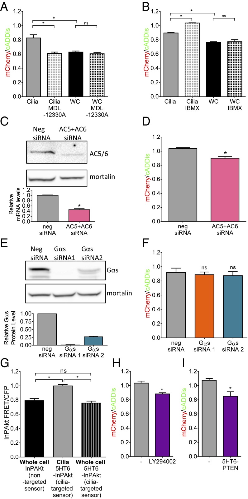
Fig. 4.
(A) Direct inhibition of AC using 100 μM MDL-12330A for 30 min reduces ciliary cAMP to whole-cell levels (*P < 0.05; n = 3 experiments) but does not affect whole-cell cAMP levels. (B) Inhibition of PDE using 100 μM IBMX overnight increases ciliary cAMP but not whole-cell cAMP levels (*P < 0.05; n = 3 experiments). (C) Western blot using an anti-AC5/6 antibody showing effective knockdown using siRNA to AC5 and AC6; mortalin is used as a loading control. The bar graph shows relative reduction in AC6 mRNA (*P < 0.05). (D) Knockdown of AC5/6-reduced ciliary cAMP measured using the mCherry/cADDis ratio (*P < 0.05; n = 3 experiments; 15 to 40 ROIs per experiment). (E) Western blot showing effective siRNA knockdown of Gαs in MEFs; mortalin is used as a loading control. The bar graph is a quantitation of three independent knockdown experiments. (F) Knockdown of Gαs does not reduce ciliary cAMP measured using the mCherry/cADDis ratio (n = 3 experiments; 7 to 14 ROIs per experiment). (G) Summary FRET/CFP data from cells expressing the PIP3 sensor InPAkt or 5HT6-InPAkt in cilia (*P < 0.05; n = 3 experiments) or the whole cell (n = 3 experiments) show that cilia have higher PIP3 levels. (H) Inhibition of PI3K using 10 nM LY294002 overnight reduces basal cilia cAMP levels (*P < 0.05; n = 3 experiments). (I) Expression of the lipid phosphatase PTEN in cilia reduces cAMP levels (*P < 0.05; n = 3 experiments). Data are presented as mean ± SEM.

