Fig. 5.
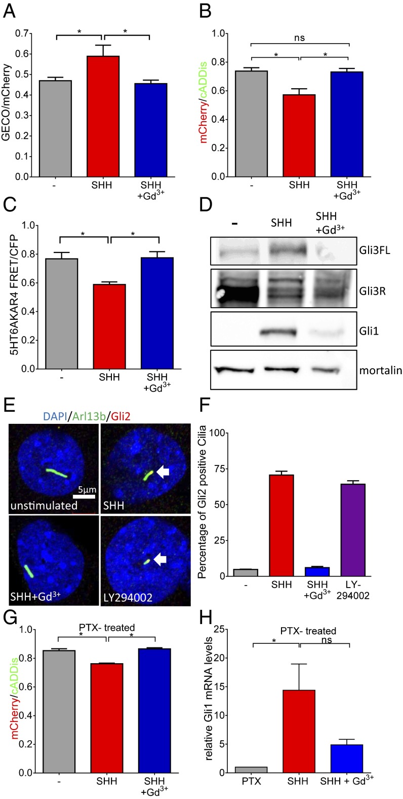
SHH effects on Ca2+, cAMP, PKA, and Gli. (A) Overnight application of 100 μM Gd3+ inhibits SHH (10 nM)-mediated increase in ciliary Ca2+ (*P < 0.05; n = 3 experiments; 7 to 12 ROIs each). (B) SHH reduces ciliary cAMP; Gd3+ prevents the effect of SHH (*P < 0.05; n = 3 experiments). (C) SHH inhibits PKA activity in cilia measured using the PKA sensor AKAR4. Gd3+ prevents SHH inhibition of PKA (*P < 0.05; n = 3 experiments). (D) In MEFs, SHH reduces Gli3R and increases Gli3-FL and Gli1; Gd3+ inhibits SHH effects. Western blot is representative of three similar experiments. Mortalin is used as a loading control. (E) Immunofluorescence to detect localization of transfected Gli2-HA in cilia after various treatments. Arl13b immunofluorescence (green) is used to demarcate cilia; nuclei are stained using DAPI. Gli2 localizes to cilia after SHH treatment. The SHH effect on Gli2 localization is inhibited by Gd3+. Inhibition of PI3K leads to localization of Gli2 to cilia in the absence of SHH. White arrows indicate colocalization. (F) Summary data for ciliary localization of Gli2-HA after various treatments. The data are from two independent experiments. A total of 42 to 55 cilia were counted for the four conditions to determine the percentages. (G) In MEFs treated with PTX (250 ng/mL; overnight), SHH maintains its ability to reduce ciliary cAMP whereas Gd3+ still prevents the SHH effect (*P < 0.05; n = 3 experiments). (H) SHH increase of Gli1 expression is also maintained after PTX treatment but was prevented by Gd3+ (*P < 0.05). Data are presented as mean ± SEM.

