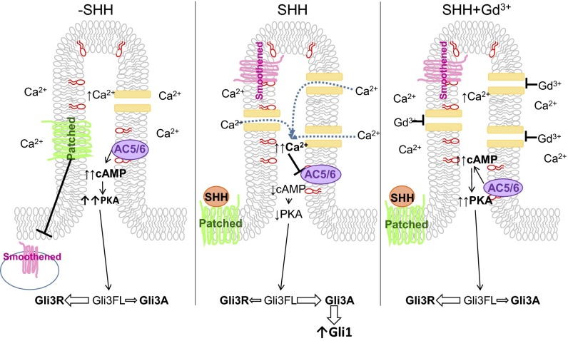
Fig. 6.
Model of ciliary HH signaling. (Left) Under basal conditions, PIP3 (depicted in red on the inner leaflet of the membrane) maintains AC5/6 activity in the cilia, resulting in high cAMP concentration. Relative to the whole cell, high Ca2+ is present in the cilia. Basal cAMP activates PKA, shifting the Gli balance in favor of GliR. (Middle) After SHH stimulation, Smoothened translocates to the ciliary membrane, and channel activity and Ca2+ levels are increased (12) that inhibit AC5/6. The resulting reduction in cAMP reduces PKA activity, shifting the balance to GliA. (Right) Gadolinium blocks Ca2+ entry through channels, inhibiting the effects of SHH.

