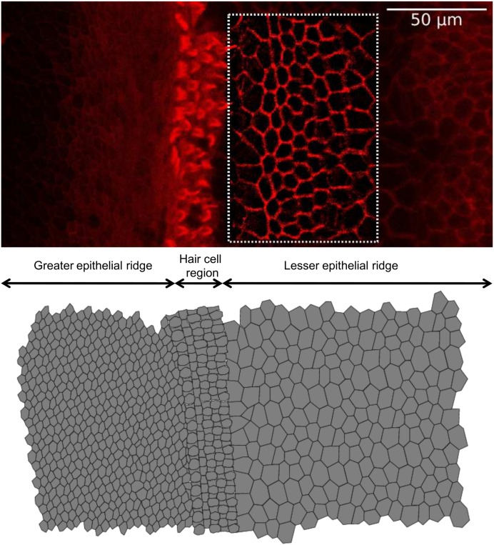
Fig. S1.
Construction of realistic tissue morphology from confocal fluorescence imaging of a cochlear organotypic culture. (Top) Representative surface view of the cochlear sensory epithelium in an organotypic culture (P5, mouse). Image contrast was enhanced by application of an unsharp mask filter and an edge detection algorithm. (Bottom) Digitally reconstructed epithelium. The set of differential equations (General Description of the Computational Model) was solved iteratively for each cell in the network and intercellular diffusion of IP3 was computed for each pair of neighboring cells. Methods: Cochlear organotypic cultures were fixed in 4% paraformaldehyde for 20 min at room temperature and rinsed in PBS containing 2% BSA. F-Actin was stained for 1 h at room temperature with Texas Red-X phalloidin (ThermoFisher Scientific, no. T7471). After being washed three times in rinse solution, samples were mounted onto glass slides with a mounting medium (FluorSave Reagent; Merk, no. 345789). Images were acquired using a laser scanning confocal microscope (SP5; Leica).

