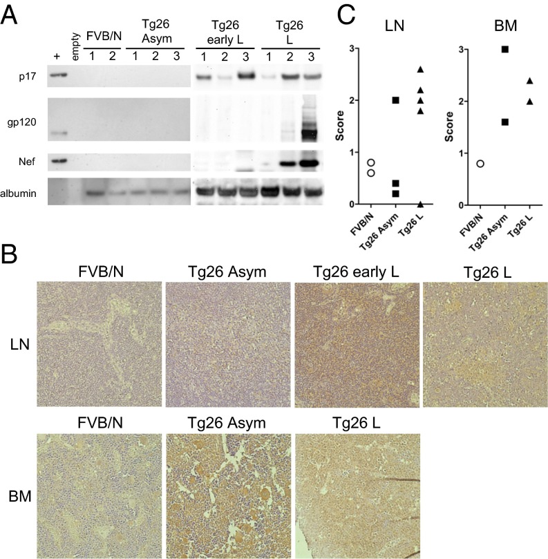
Fig. 1.
Expression of HIV proteins in lymph nodes and BM of Tg26 mice. (A) Lymph nodes from each mouse were pooled, and cell lysates were probed by Western blot analysis for HIV proteins p17, gp120, Nef, or albumin as a loading control. (B) IHC for p17 in lymph nodes (Top) and BM (Bottom) of Tg26 mice. Positive staining is indicated by deposition of DAB (brown); tissues were counterstained with hematoxylin. As a negative control, FVB/N tissues displayed little background staining (Left). (Original magnification 400×.) (C) Scoring of p17 staining as described in Materials and Methods. Each symbol represents the mean score of one section per mouse.

