Fig. 2.
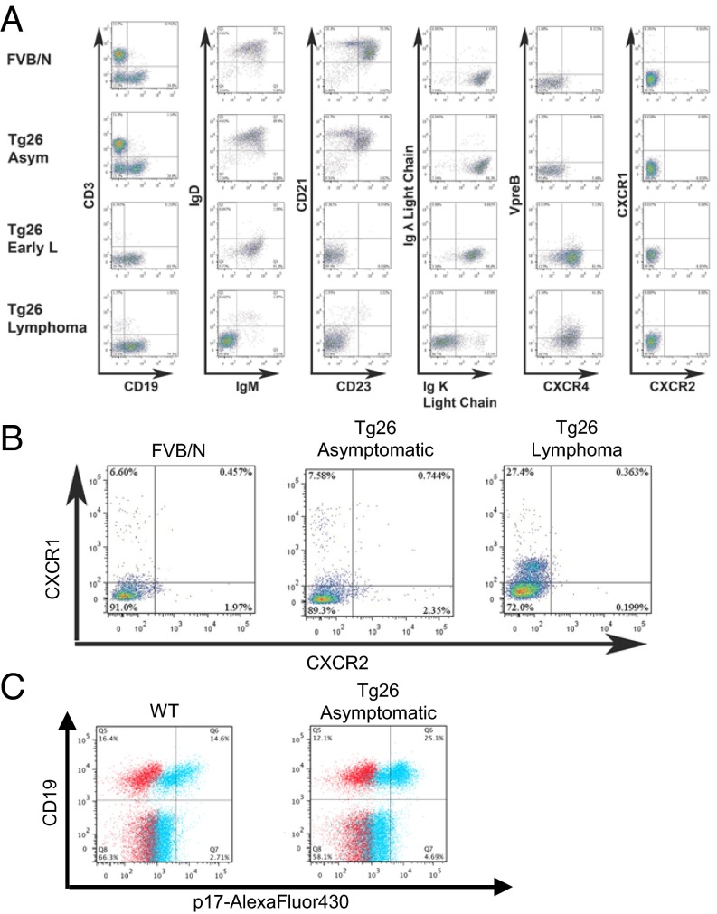
Characterization of peripheral B cells in Tg26 mice. (A) Spleen cells from the indicated mice were stained for surface markers and analyzed by flow cytometry. Plots are gated on B cells (CD3−CD19+) shown at left. Data are representative of three mice per group. (B) Mouse PBMCs were gated on B cells as in A and analyzed for CXCR1 and CXCR2 expression. (C) PBMCs from WT and Tg26 mice were analyzed for binding of p17-Alexa Fluor 430. Live PBMCs were gated on exclusion of Live/Dead dye and low SSC profile (not shown). Blue: stained with p17-Alexa Fluor 430; red: stained without p17-Alexa Fluor 430. A majority of CD19+ B cells bound labeled p17, whereas CD19− cells showed minimal binding.

