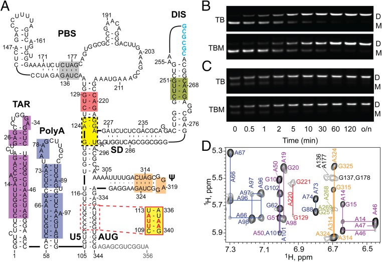
Fig. 1.
(A) Secondary structure of the HIV-1 5′-L356 in its DIS-exposed, dimer-promoting state (adapted from ref. 57; residues truncated in 5′-L344 colored gray). The 5′-L344 NMR signals with resolved and assigned 2D 1H–1H NOESY cross-peaks are denoted by color shaded boxes; yellow denotes sites that were only assignable in mutant constructs that either lacked the upper PBS loop (5′-L344ΔPBS) (57) or contained an lrAID substitution in the U5:AUG stem (5′-L344-UUA) (48). Intact (B) and truncated (C) 5′-leader constructs rapidly dimerize when incubated in PI buffer at 37 °C. Dimerization profiles on TB and TBM gels are indistinguishable, indicating that the dimers are nonlabile. o/n, overnight. (D) Region of the 2D 1H–1H NOESY spectrum of A2rGrCr-labeled 5′-L344 RNA. Assignments (adenosine-H2 and ribose-H1′ assignments labeled vertically and horizontally, respectively) are color-coded to match the highlighted elements within the secondary structure in A.

