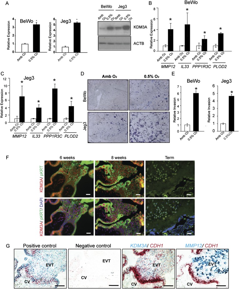
Fig. S5.
Hypoxia responses of human trophoblast cells and protein and transcript localization in human placental tissues. (A) Effects of low oxygen (0.5% O2) on KDM3A transcript (qRT-PCR) and protein (Western blot) expression in BeWo and Jeg3 human trophoblast cells. Amb, ambient. (B and C) Effects of 0.5% O2 on MMP12, IL33, PPP1R3C, and PLOD2 expression in BeWo (B) and Jeg3 (C) human trophoblast cells. Statistical analysis for A–C: n = 3/group; Mann–Whitney test, *P < 0.0.5. (D) Effects of 0.5% O2 on invasive behavior of BeWo and Jeg3 human trophoblast cells. Images are representative Insets. (E) Quantification of invasion through Matrigel (Student t test, *P < 0.05). (F) First trimester (6 and 8 wk) and term human placental tissues were processed for immunohistochemistry using KDM3A and pan cytokeratin (pKRT) antibodies. (Scale bar, 25 μm.) (G) In situ hybridization analysis of first trimester human placental tissue, including positive [POLR2A (red) and PPIB (blue)] and negative (DapB) controls, and colocalization of KDM3A (blue, third image) and MMP12 (blue, fourth image) with CDH1 (red). (Scale bar, 100 μm.)

