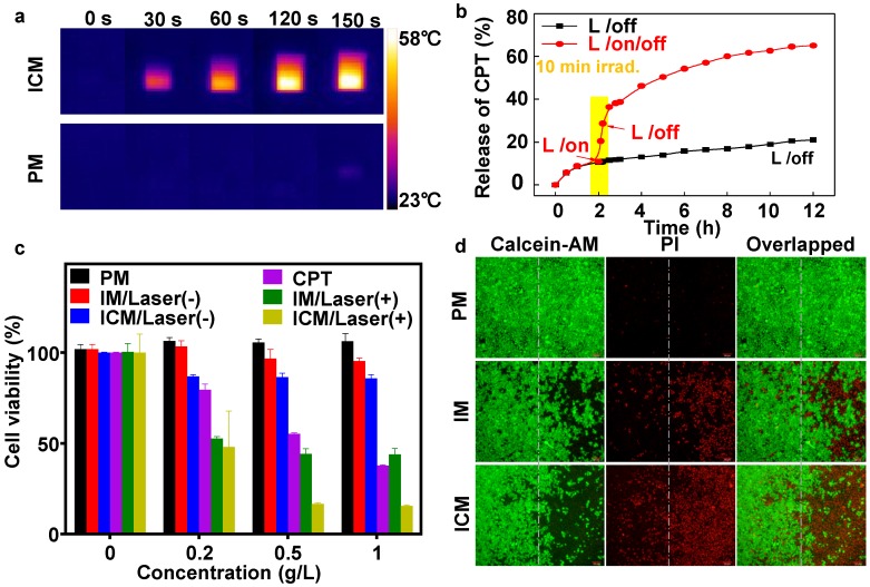
Figure 3.
(A) NIR thermal imaging of ICM and PM after treated by 808 nm laser at power density of 1 W cm-2 for 150 seconds. (B) The CPT release profile of ICM with and without laser irradiation (808 nm, 1 W cm-2) for 12 h. (C) The viability of HeLa cells treated with (+; for 10 min) or without (-) laser irradiation after being incubated with various micelles and concentrations. The error bars are based on the standard deviations of five parallel samples. (D) Fluorescence images of calcein AM/PI co-stained HeLa cells after incubation with PM, IM, and ICM upon being exposed to 808 nm laser at power density of 1 W cm-2 for 10 min (right side). Cells incubated with the same concentration (0.5 g/L) of polymeric micelles without laser irradiation were chosen as controls (left side).

