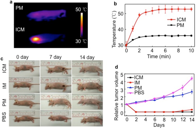
Figure 6.
(A) In vivo NIR thermal imaging obtained 24 h after the intravenous injection of PM or ICM (200 µL, 2.0 g/L) into mice and treatment by an 808 nm laser at a power density of 1 W cm-2 for 10 min. (B) Temperature change at the tumor sites as a function of irradiation time. (C) Effects of photothermal therapy in HeLa tumor-bearing mice. Representative time-dependent photos taken for mice after being irradiated by 808 nm NIR light with ICM, IM, PM (200 µL, 2.0 g/L), and PBS post-injection. (D) Tumor volumes of different groups measured after laser irradiation and normalized to their initial size (n = 5 per group). Error bars indicated the means and standard errors.

