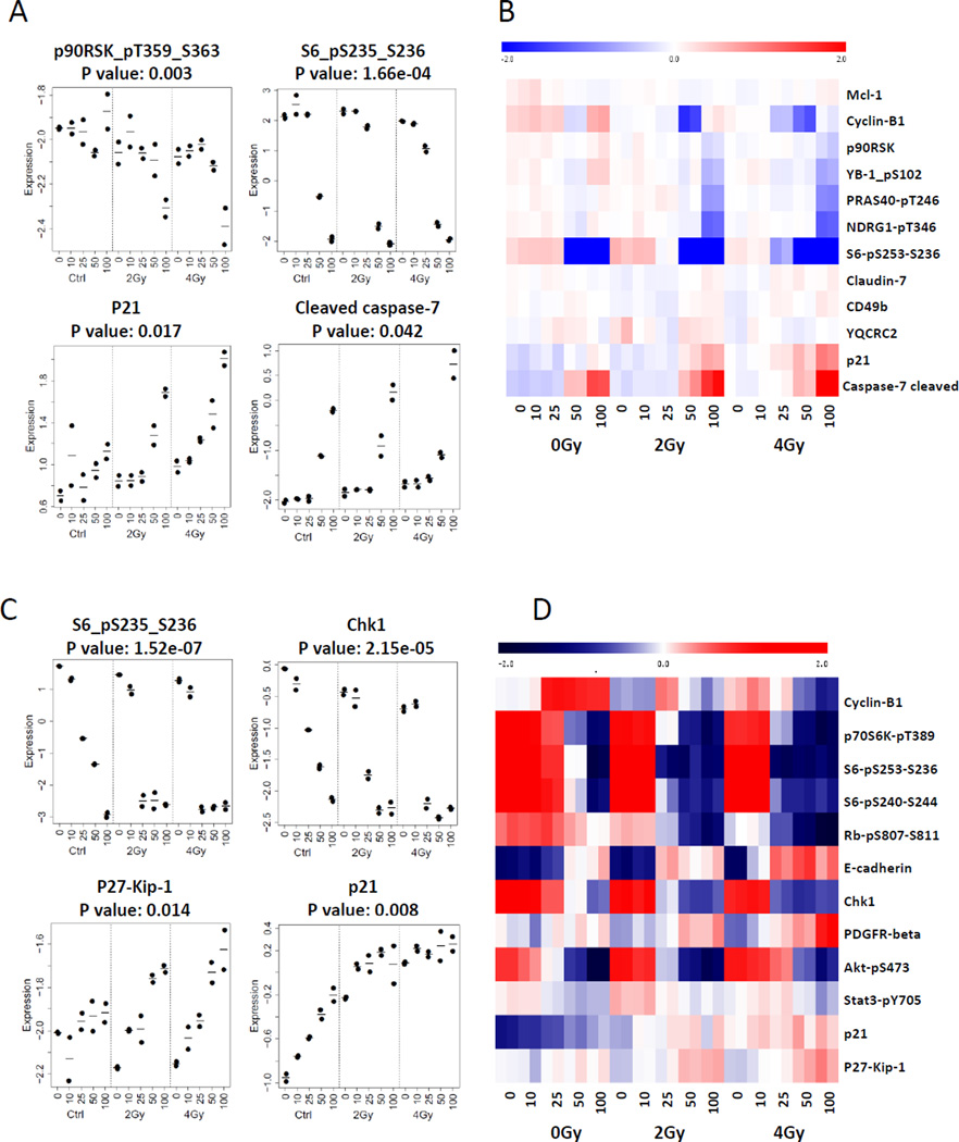
Figure 4. Ganetespib altered expression level and activity of client growth factors and cell cycle progression proteins.
H460 and A549 cells were treated with ganetespib at 0 nM, 10 nM, 25 nM, 50 nM and 100 nM 5 hrs prior to irradiation (0 Gy, 2 Gy and 4 Gy) followed by an additional 19 hours of post-irradiation incubation in ganetespib containing medium. H460 and A549 cell lysates were collected for RPPA analysis (A–D). Heat map of RPPA results indicated altered activities or expression level of proteins (B and D).

