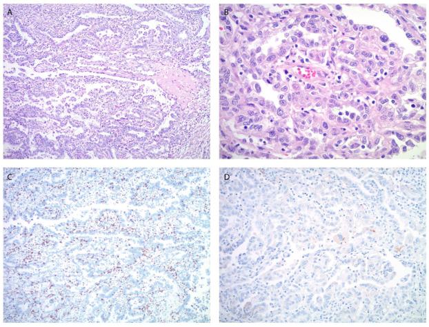
Figure 1.
Microscopic images of the endometrial carcinoma in patient #1. A, B. The tumor shows predominantly a papillary and glandular architecture (A) with high nuclear grade and clear to pale eosinophilic cytoplasm (B), consistent with clear cell carcinoma component. C. Moderate peri- and intratumoral lymphocytic infiltrate is highlighted by CD8 immunostain. D. PD-L1 immunostain shows focal, weak staining predominantly located in the tumor cells. (A. Original magnification 100x, H&E stain, B. Original magnification 400x, H&E stain, C. Original magnification 100x, D. Original magnification 200x).

