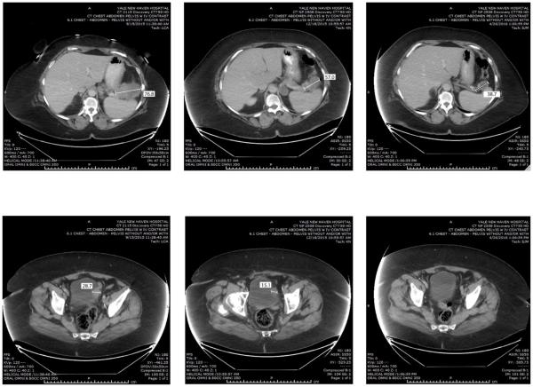
Figure 2.
Representative CAT scans demonstrating activity (ie, partial response) of nivolumab in Patient #1 (ie, POLE-ultra-mutated). Left panels: Pretreatment images with baseline measurements of two representative metastatic tumor deposits [ie, mass abutting the pancreatic tail in the gastro-splenic ligament (Upper Left Panel) and mass involving the bladder dome wall (Lower Left Panel). Middle panels: Regression of the metastatic tumor deposits described above three months after treatment initiation. Right panels: regression (Upper Right) and disappearance (Lower Right) of the metastatic tumor deposits described above seven months after treatment initiation with nivolumab.

