
Figure 2. JAK2V617F vascular niche contributes to the growth advantage of JAK2V617F HSPC over JAK2WT HSPC.
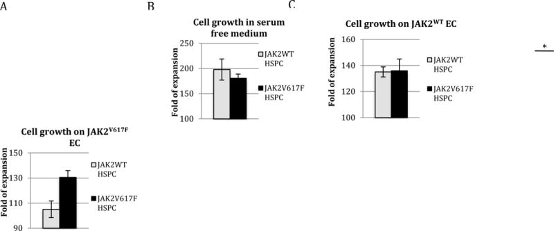
JAK2WT (from control mice) and JAK2V617F (from Tie2/FF1 mice) Lin−cKit+ HSPCs were cultured on a feeder layer of JAK2WT or JAK2V617F ECs under serum-free conditions. (A) There was no significant difference between JAK2WT and JAK2V617F HSPC proliferation in vitro in SFEM. (B) There was no significant difference between JAK2WT and JAK2V617F HSPC proliferation when co-cultured with JAK2WT EC. (C) JAK2V617F HSPC displayed a relative growth advantage over the JAK2WT HSPC when co-cultured with JAK2V617F EC. Cell proliferation was shown as fold of expansion which is the ratio of the final cell count to starting cell count. The results were expressed as mean ±s.e.m. (n=3) Data are from one of two independent experiments (with triplicates in each experiment) performed by two investigators (C.L. and H.Z.) that gave similar results. * p < 0.05