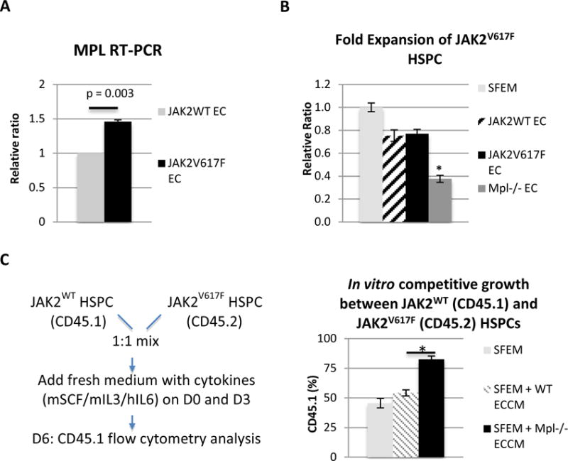
Figure 3. MPL receptor on vascular ECs contributes to JAK2V617F HSPC maintenance/expansion in MPN.

(A) MPL expression was increased by 46% in the JAK2V617F ECs compared to JAK2WT ECs (p = 0.003). MPL expression in JAK2V617F ECs was shown as the relative ratio compared to its expression in JAK2WT ECs which was set as “1”. (B) JAK2V617F HSPC (Lin-cKit+) cell proliferation was significantly decreased when co-cultured with MPL−/− EC, compared to when co-cultured with JAK2WT ECs or JAK2V617F ECs. Cell proliferation in EC co-culture was shown as the relative ratio compared to cell proliferation in SFEM which was set as “1”. (C) Left: experimental design of an in vitro competitive growth assay where JAK2WT HSPCs (CD45.1) and JAK2V617F HSPCs (CD45.2) were cultured together (1:1 mix) in the presence of ECCM collected from either WT EC or MPL−/− EC. Right: while there were equal numbers of JAK2WT HSPCs (CD45.1) and JAK2V617F HSPCs (CD45.2) in the presence of WT ECCM, there were significantly more JAK2WT HSPCs than JAK2V617F HSPCs in the presence of MPL−/− ECCM. The results were expressed as mean±s.e.m. (n=3). Data were from one of two independent experiments (with triplicates in each experiment) performed by two investigators (C.L. and H.Z.) that gave similar results.
