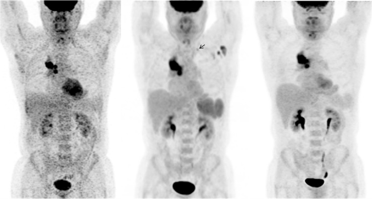
Fig. 1.
Initial staging 18F-fluorodeoxyglucose (FDG)-positron emission tomography (PET)/computed tomography (CT) was performed in a 55-year-old man with right lung cancer. Maximum intensity projection (MIP) images show the metabolically active right lung mass and ipsilateral hilar metastases, but also new enlarging FDG-avid left axillary lymph nodes, a left supraclavicular lymph node, and intense splenic FDG uptake (middle) compared to an FDG-PET/CT scan performed at an outside institution two months prior (left). The splenic uptake raised the question of a systemic inflammatory process and further questioning revealed that the patient received an influenza vaccine in the left upper extremity 2–3 days before the examination (middle). Since contralateral metastases would alter management, FDG-PET/CT was performed 12 days later without interval therapy (right). Left axillary and supraclavicular nodal and splenic FDG uptake resolved and was presumably due to local and systemic immune-mediated responses to the vaccine

