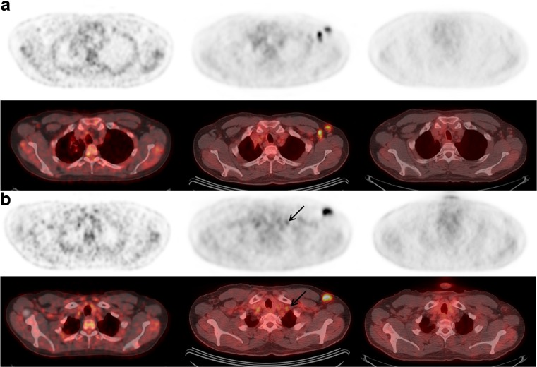
Fig. 2.
Axial PET (a and b, top rows) and fused PET/CT (a and b, bottom rows) images from FDG-PET/CT examinations performed before (left), 2–3 days following (middle), and 12 days following the influenza vaccination (right) administered in the left upper extremity. Compared to the initial scan (left), new enlarged and FDG-avid left axillary lymph nodes (a) were seen on the examination 2–3 days following the influenza vaccination (middle), which resolved 12 days later (right). Several previous reports demonstrated the occurrence of false-positive ipsilateral FDG-avid axillary lymph nodes following vaccination [1–4]. The highest uptake typically occurred within the first 2 weeks after vaccination [1, 3], but persisted beyond 1 month in some cases [2]. Additionally, lymph node size was normal [1–4], in contrast to our case. These reports advocate caution when considering changes to the treatment plan prior to confirmation of the suspected false-positive uptake. FDG uptake can also be seen in lymph nodes ipsilateral to an FDG injection site, especially if tracer is extravasated. In this case, FDG was injected in the left antecubital fossa 2–3 days after vaccination, and in the right hand approximately 2 weeks later. This is a confounding factor in this case; however, the ipsilateral FDG injection alone does not explain the enlarging size of the left axillary lymph nodes, the left supraclavicular lymph node (arrows, b and Fig. 1) and the increased splenic uptake compared to the outside scan before vaccination (left, Fig. 3)

