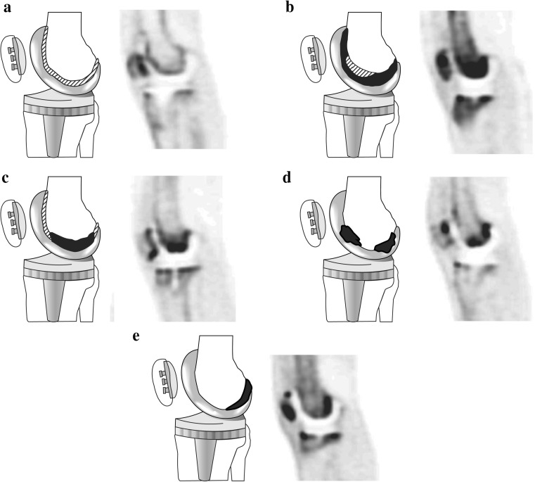
Fig. 4.
Diagrams and representative PET images showing the five types of uptake pattern in the femoral component of knee prostheses (a type 1, b type 2,. c type 3, d type 4, e type 5) Black areas represent areas of severely increased uptake and shaded areas represent areas of mildly increased uptake

