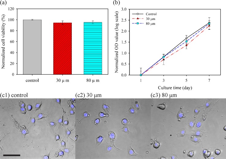
FIG. 6.
Effects of nozzle diameter on viability, proliferation, and morphology of NIH/3T3 cells. (a) Normalized cell viability after pipetting for the control group and each printing through a live/dead assay kit. (b) Assessment of proliferation through the normalized optical density (OD) value at 450 nm by each OD value at day 1, corresponding to initial population with CCK-8 assay. Slopes of curves imply the growth rate. (c) Cell morphology and fluorescence of nuclei stained by Hoechst 33342 with cultured cells at day 5 (Scale bar = 50 μm).

