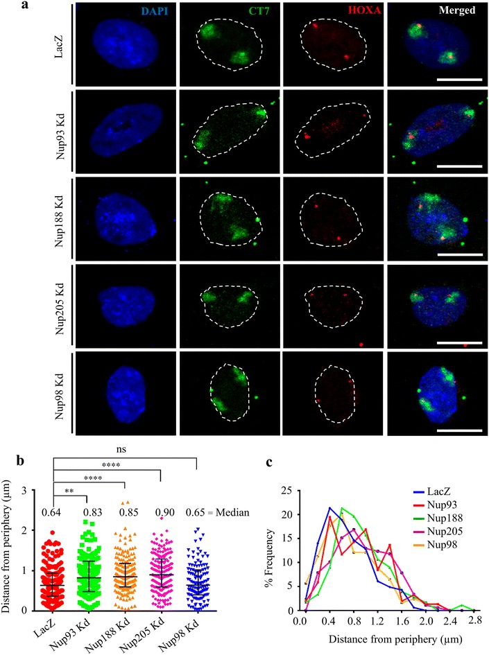
Fig. 4.

HOXA gene loci is untethered from the nuclear periphery upon Nup93, Nup188 and Nup205 depletion. a Representative images (maximum intensity projection of a confocal image stack) of 3D-FISH for HOXA (red), CT7 (green) and DAPI (blue) performed on siLacZ-, Nup93-, Nup188-, Nup205- and Nup98-depleted DLD1 cells. Scale bar ~10 μm, white dotted line indicates nuclear boundary. b Dot scatter plot showing shortest distance of HOXA gene locus from nuclear periphery demarcated by DAPI in siLacZ (n = 164 loci signals)-, Nup93 (n = 154)-, Nup188 (n = 178)-, Nup205 (n = 178)- and Nup98 (n = 124)-depleted DLD1 cells, horizontal bar represents median with interquartile range. Data from two independent biological replicates, **p < 0.01; ****p < 0.001 (Kolmogorov–Smirnov test). c % Frequency distribution profile of HOXA gene locus from nuclear periphery plotted as bins of ~0.2 µm each from the nuclear periphery. Y-axis represents % frequency of HOXA locus pooled from two independent biological replicates
