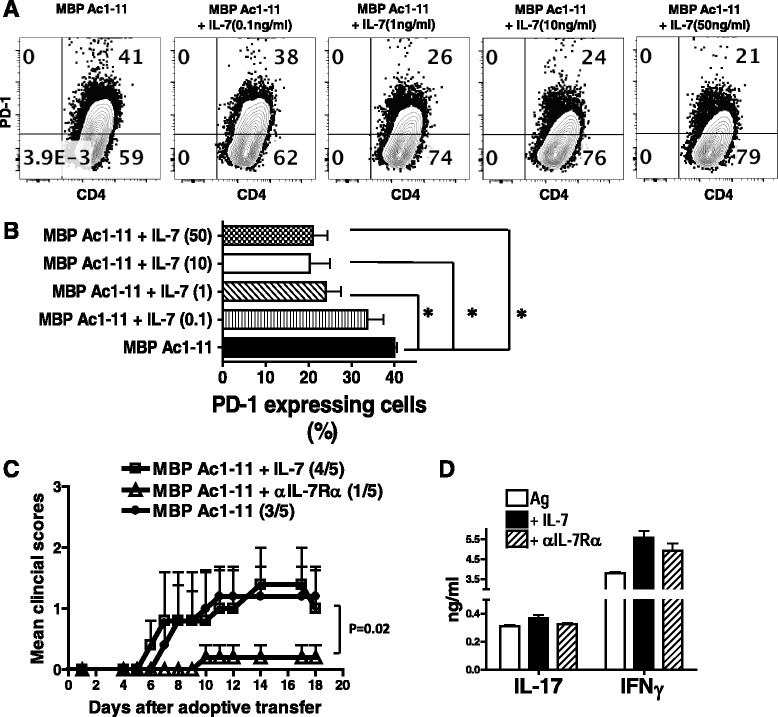
Fig. 5.

IL-7 inhibits PD-1 expression in myelin-specific CD4 T cells. a Splenocytes from naive TCRβ transgenic mice were activated with MBP Ac1-11 plus different concentration of rmIL-7 for 6 days. PD-1 expression was determined by flow cytometry. b The percentage of PD-1 expressing cells in the groups with IL-7 (concentrations as indicated) was compared to that in MBP Ac1-11 only group. Cells were gated on CD4+ CD44+ cells and flow data are representative of three independent experiments. Error bars denote s.e.m. *P < 0.05. c Splenocytes from naive TCR-WT mice were activated with MBP Ac1-11, MBP Ac1-11 plus rmIL-7 (10 ng/ml), or MBP Ac1-11 plus αIL-7Rα (0.5 μg/ml) for 3 days and transferred into naive B10 PL recipient mice by intraperitoneal (i.p.) injection. The mice were monitored for EAE development. d IFNγ and IL-17 in supernatant were determined by ELISA. Disease incidence (sick mice/total mice) is indicated in parentheses. Data are representative of two independent experiments
