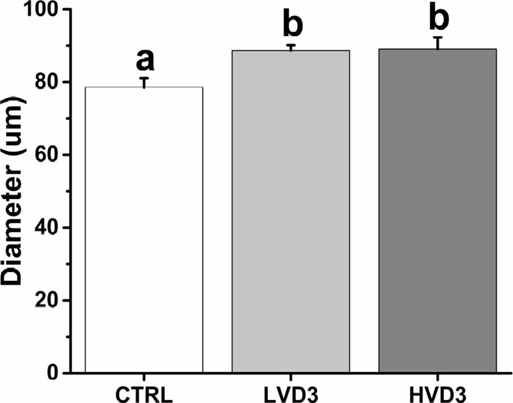
Figure 4.
The effects of vitamin D3 on oocyte growth in rhesus macaque antral follicles after 5 weeks of culture in an alginate matrix. Oocyte growth was determined by measuring oocyte diameters. CTRL, control; LVD3, low-dose (25 pg/ml) vitamin D3 addition; HVD3, high-dose (100 pg/ml) vitamin D3 addition. Significant differences between treatment groups are indicated by different letters (P < 0.05). Data are presented as the mean ± SEM with 14–28 oocytes per experimental group.

