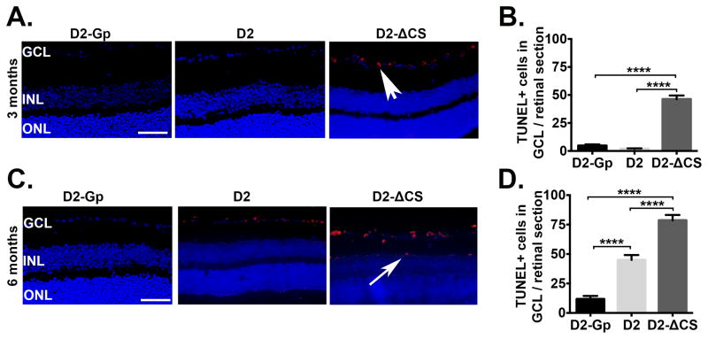
Fig 2. Accelerated apoptosis coincides with RGC loss in the absence of sFasL.
Representative TUNEL staining in paraffin embedded retinal sections taken from D2-Gp, D2 and D2-ΔCS mice at (A) 3 and (C) 6 months of age (Scale bar, 100μm). TUNEL = red, DAPI, nuclear marker = blue. GCL, ganglion cells layer; INL, inner nuclear layer; ONL, outer nuclear layer. White arrowhead = TUNEL positive cells in GCL, white arrow = TUNEL positive cells in INL. TUNEL positive cells in the GCL were quantitated at (B) 3 months and (D) 6 months of age, shown as the number of TUNEL positive cells in the GCL/retinal section (9 sections/retina), N=8 per group. ****P<0.0001.

